Presentation
Abnormal CT chest and hence MRI of heart ordered for evaluating cardiac apex.
Patient Data
Note: This case has been tagged as "legacy" as it no longer meets image preparation and/or other case publication guidelines.
![](https://prod-images-static.radiopaedia.org/images/17675525/4f471a969e5f04452409ea24a499ab_thumb.jpeg)
![This study is a stack](/packs/stack-YQKLCKBI.gif)
![](https://prod-images-static.radiopaedia.org/images/17675610/c9a496150ca334c0e42bceadc565f7_thumb.jpeg)
![This study is a stack](/packs/stack-YQKLCKBI.gif)
![](https://prod-images-static.radiopaedia.org/images/17675525/4f471a969e5f04452409ea24a499ab_big_gallery.jpeg)
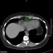
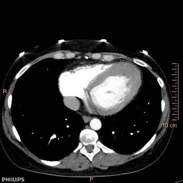
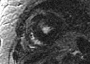
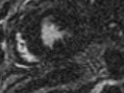
Fat density seen at apex of the heart.
Apex is thick, easier to appreciate in retrospect.
![](https://prod-images-static.radiopaedia.org/images/18117843/b129b77e4c86718d0f70efff1eb997_thumb.jpeg)
![](https://prod-images-static.radiopaedia.org/images/18117844/15107aa64c7f3f06351888dd2a722c_thumb.jpeg)
![](https://prod-images-static.radiopaedia.org/images/18117845/5125913b461b6028d089ecae325f93_thumb.jpeg)
![](https://prod-images-static.radiopaedia.org/images/18117846/57afc2912410da3039d7cdeffb6578_thumb.jpeg)
![](https://prod-images-static.radiopaedia.org/images/18117847/9c41aea2be661be7b4e794d35d0d27_thumb.jpeg)
![](https://prod-images-static.radiopaedia.org/images/18117848/2dd836c82cad6b43c231e111cba76e_thumb.jpeg)
![](https://prod-images-static.radiopaedia.org/images/18117849/5eca8e6a1afdf568f0e9600743d91b_thumb.jpeg)
![](https://prod-images-static.radiopaedia.org/images/18117850/91117ea65c3d59d4fb5b7e1a81bdf5_thumb.jpeg)
![](https://prod-images-static.radiopaedia.org/images/18117851/a833e1b0bfc0c88b71c2fbaabda946_thumb.jpeg)
![](/packs/icon_video_small-FOK545SU.png)
![](https://prod-images-static.radiopaedia.org/images/18117843/b129b77e4c86718d0f70efff1eb997_big_gallery.jpeg)
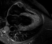
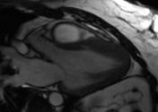
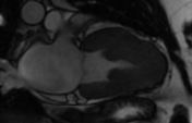
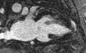
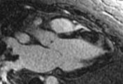
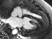
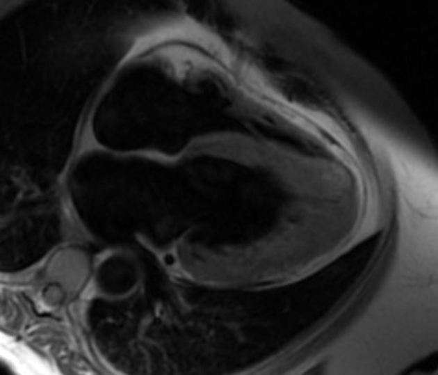
Left ventricular function: hyperdynamic left ventricular systolic function
Valvular function: no systolic anterior motion of the mitral valve
Delayed enhancement: Tiny focus of late gadolinium enhancement at the distal apex of the septum towards the left ventricle
Case Discussion
Findings are suggestive of apical hypertrophic cardiomyopathy. There is no apical aneurysm.
Recommend a 12-lead electrocardiogram to evaluate for T-wave inversion. Look at the apex on the 3-chamber view in diastole, it is not thinned out as usual. It has a spade-like appearance.