
Presentation
Increasing abdominal pain and fever
Patient Data
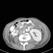

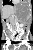

Very large multilobulated, septated, calcified cystic left splenic lesion is again seen consistent with the known hydatid cyst measuring 21.5 x 14.6 x 21.8 cm, displacing the stomach to the right, and the left kidney, splenic flexure and pancreatic tail inferiorly. It is predominantly filled with low density fluid, but also contains a moderate volume of gas laterally. No free intraperitoneal gas is seen. A large volume of free intraperitoneal fluid is seen. The liver is surrounded by free fluid and has an extremely nodular surface with scalloping of the capsule. Hypodense lesions in segment 6 and 8 are stable. Tiny segment 5 calcific granuloma. Stable hyperdense right renal cyst. The remainder of the intra-abdominal and pelvic organs are unremarkable.
Large left pleural effusion.
Conclusion
- Very large splenic hydatid cyst with free intraperitoneal fluid consistent with cyst rupture. New intra-cystic gas of uncertain source and may represent secondary infection.
- Stable liver cystic lesions.
Case Discussion
The scalloped nature of the liver is typical for the thick fluid released from the ruptured hydatid cyst.


 Unable to process the form. Check for errors and try again.