
Presentation
Seizure.
Patient Data
Note: This case has been tagged as "legacy" as it no longer meets image preparation and/or other case publication guidelines.
ASNR 2016: This case was submitted as part of the American Society of Neuroradiology (ASNR 2016) Case Of The Day competition, in collaboration with Radiopaedia.org.
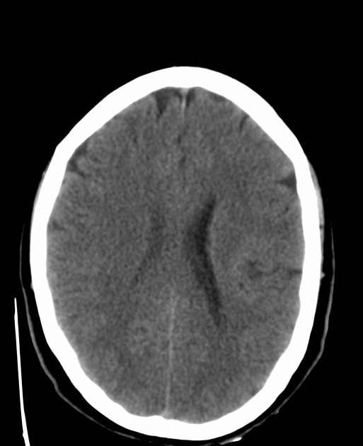
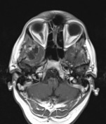
Non-contrast axial brain CT scan shows a cystic lesion in the right temporal lobe. Hyperdense areas are seen in the tumor periphery that may be due to hemorrhage or a solid component.
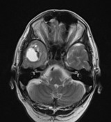
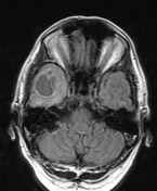
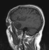
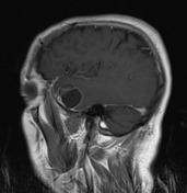

There is a solid-cystic mass in the right temporal lobe. The internal signal is not completely suppressed in FLAIR images. A finding that suggests high protein contents of the cyst. The adjacent edema is minimal. Solid mural nodule shows enhancement after contrast administration.
Case Discussion
The histologic examination confirmed pleomorphic xanthoastrocytoma (PXA).
PXAs are solid-cystic variants of WHO class II astrocytomas that most commonly appear in the temporal lobe of adult patients. The solid component enhances avidly after contrast administration. The adjacent edema is minimal.


 Unable to process the form. Check for errors and try again.