
Chronic distal ileal obstruction due to a polyp: neuroendocrine tumor
Presentation
2 years of intermittent abdominal pain and distension. Treated as irritable bowel syndrome. Symptoms persisting.
Patient Data
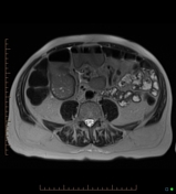
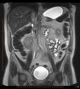
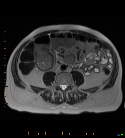
CT scan (two years ago)
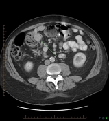

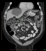
Distension and faecalization of distal small bowel with abrupt transition to collapsed ileum just proximal to the ileo-cecal junction (arrow). Note more proximal small bowel is normal. The distal ileum has an appearance similar to the adjacent transverse colon. Findings not appreciated by the reporting radiologist thus no surgical treatment instituted.
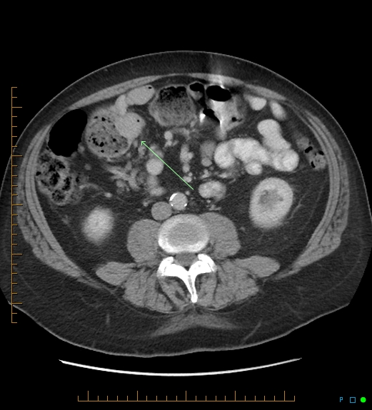
MRI examination (current)
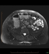
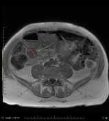
Rounded mass (arrows) in the distal ileum with faecalization and dilatation of ileum proximal to the mass and collapse distally. Mass is of relatively low signal intensity on T1 and T2 thus not a lipoma. Has been present for over 2 years thus not malignant. Incidental gallbladder calculi and multiple cysts (liver and kidney).
Case Discussion
This case demonstrated the so-called faecalization of small bowel content ie small bowel lumen containing material that is more akin to the colon rather than the normal fluid-like appearance. It is an important CT sign as it is never normal thus indicating the presence and chronicity of small bowel obstruction.
At surgery, this case was confirmed to be a low-grade neuroendocrine tumor.


 Unable to process the form. Check for errors and try again.