
Presentation
Abdominal pain. Blood results show high lactate, low hemoglobin, and acidosis.
Patient Data


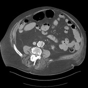


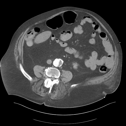
Bleeding peptic ulcer in the duodenal bulb. Associated bowel wall thickening and fat stranding. No free gas or extraluminal (i.e. peritoneal) hemorrhage. On portal venous phase imaging there is pooling of contrast in the distal duodenum and proximal jejunum.
Most likely bleeding point is the gastroduodenal artery arising from the left hepatic artery. Replaced right hepatic artery is noted. 4 cm infrarenal abdominal aortic aneurysm with mural thrombus.
Inferior midline ventral hernia containing non-obstructed large and small bowel, and omental fat.
Case Discussion
Peptic ulcers are the most common cause of non-variceal upper gastrointestinal bleeding. Other common causes include erosive gastritis, Mallory-Weiss tears, reflux esophagitis, and angiodysplasia.


 Unable to process the form. Check for errors and try again.