

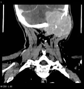
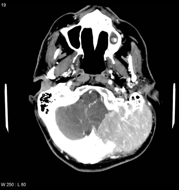
A large destructive enhancing mass arises from the left side of the occipital bone and mastoid extending into the posterior fossa.
The patient went on to have a biopsy.
Histology
Microscopic Description:
The biopsy of retroauricular mass shows skeletal muscle which is heavily infiltrated by plasma cells. While many of these are almost normal appearing, there are several extremely atypical cells with enlarged nuclei, prominent nucleoli and an increased nuclear-to-cytoplasmic ratio. The biopsy is crushed and little material was left on the immunoperoxidase slides, but these appear to show that the lesion stains for IgG kappa.
Final Diagnosis: plasmacytoma.
The patient went on to receive radiotherapy.
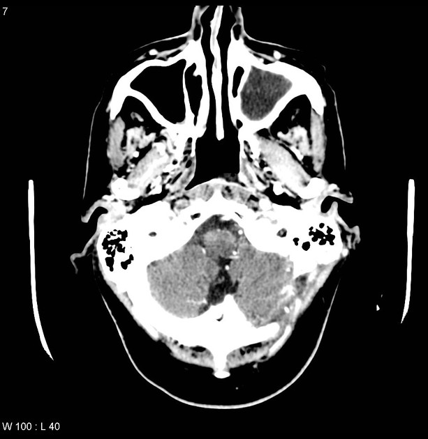
Radiotherapy resulted in marked reduction in the size of the mass and resolution of mass effect upon the posterior fossa content.
Case Discussion
The differential of masses appearing to arise from the skull includes primary bone tumors (e.g. osteosarcoma), metastases and multiple myeloma, as well as involvement by masses from within the intracranial cavity (e.g. meningioma and hemangiopericytoma) or from the scalp (e.g. squamous cell carcinoma).


 Unable to process the form. Check for errors and try again.