Presentation
Known to have "multiple hemangiomas", but appear to be growing on serial imaging exams. Relatively asymptomatic, but with slowly worsening liver function tests.
Patient Data
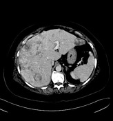

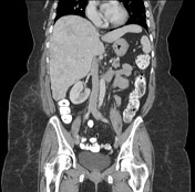

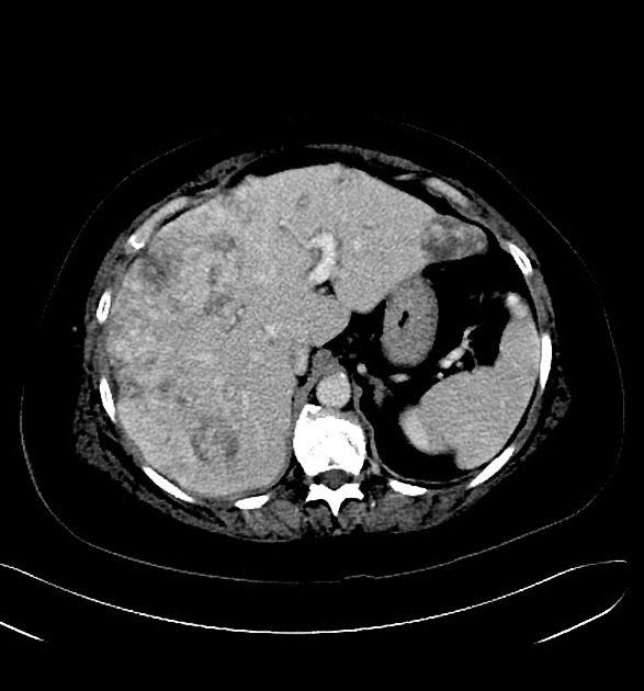
Diffuse, confluent lesions involving both the left and right hepatic lobes with enhancement on the portal venous phase. The lesions result in enlargement of the liver.
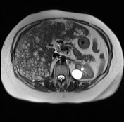

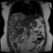

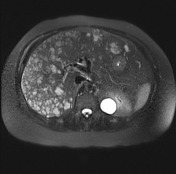

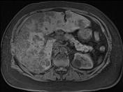


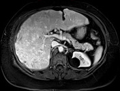

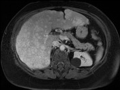

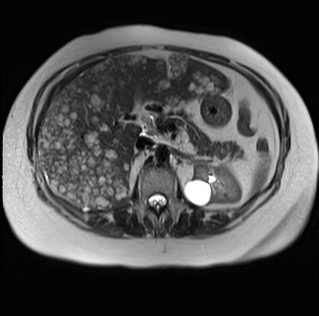
There are multiple irregular T2 hyperintense lesions in the left and right hepatic lobes, becoming confluent in the right hepatic lobe.
They are T1 hypointense with early arterial enhancement and progressive enhancement on the delayed phases.
The lesions are clustered more toward the periphery and there is mild capsular retraction.
Case Discussion
Biopsies showed atypical vascular tumor consistent with hepatic epithelioid hemangioendothelioma with focal epitheloid features. CD31+, CD34-, AE 1/3- .
Due to disease in both lobes of the liver, the patient underwent liver transplantation.


 Unable to process the form. Check for errors and try again.