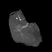
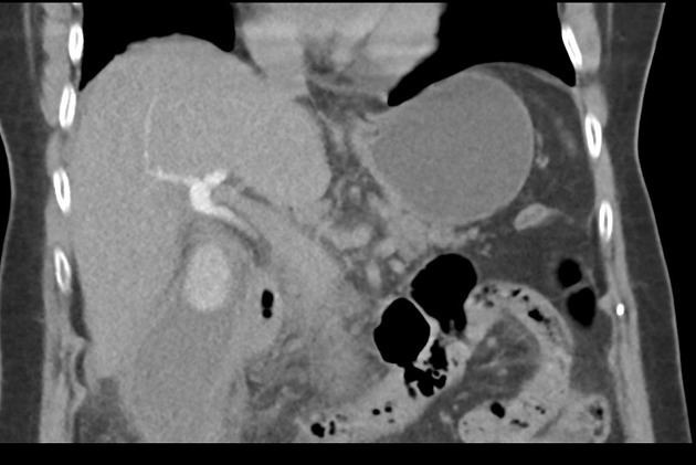
Cholecystitis - obstructive choledocholitiasis (CT intravenous cholangiography)
Presentation
Upper abdominal pain - cholecystitis + stone on external ultrasound.
Patient Data
CT IVC
![](https://prod-images-static.radiopaedia.org/images/20959484/251d0ff1c364199e2eddfe46258252_thumb.jpeg)
![This study is a stack](/packs/stack-YQKLCKBI.gif)
![](https://prod-images-static.radiopaedia.org/images/20960329/bc684c83a5ee1d534cbf7c5972accb_thumb.jpeg)
![This study is a stack](/packs/stack-YQKLCKBI.gif)
![](https://prod-images-static.radiopaedia.org/images/20961944/3768fe7fb6050736d4c7236b2d881d_thumb.jpeg)
![This study is a stack](/packs/stack-YQKLCKBI.gif)
![](https://prod-images-static.radiopaedia.org/images/20962571/627b1ab4d558544dfcc72f0c37a921baf4f7b044309d5085ea021c15e39a7134_thumb.jpeg)
![This study is a stack](/packs/stack-YQKLCKBI.gif)
![](https://prod-images-static.radiopaedia.org/images/20962446/fc37829bf9b62e7352557c2903d673f7e2e5c835581bc13ced56a9b399aabe43_thumb.jpeg)
![This study is a stack](/packs/stack-YQKLCKBI.gif)
![](https://prod-images-static.radiopaedia.org/images/20962772/a931e2dde2734691dc1f59ef9957cb587739ff8b3618aa554d769c5e267907bc_thumb.jpeg)
![This study is a stack](/packs/stack-YQKLCKBI.gif)
![](https://prod-images-static.radiopaedia.org/images/20963075/6d3c1da52d02499a6d45212d33809b59fe3803489f9e4c5bdc7f9257250aa743_thumb.jpeg)
![This study is a stack](/packs/stack-YQKLCKBI.gif)
![](https://prod-images-static.radiopaedia.org/images/20959484/251d0ff1c364199e2eddfe46258252_big_gallery.jpeg)
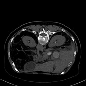
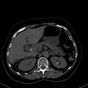
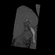
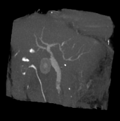
The gallbladder is distended, showing a 2.6 cm calcified stone within, thick walls, and surrounding fat-stranding and free fluid. The common biliary duct is enlarged in caliber (11mm) and demonstrates ill defined filling defects within its distal portions (measuring up to 8 mm on axial plane).
Within the limitations of a non-contrast study the remainder of the imaged upper abdominal content appears unremarkable.
Conclusion: Choledocholithiasis associated with cholecystitis.
Case Discussion
This case illustrates how the CT cholangiography can be useful in identifying choledocholithiasis. This patient has typical CT findings of cholecystitis: thick-walled and distended gallbladder with stones within, as well as surrounding inflammatory fat stranding.