
Cholecystitis - obstructive choledocholitiasis (CT intravenous cholangiography)
Presentation
Upper abdominal pain - cholecystitis + stone on external ultrasound.
Patient Data
CT IVC


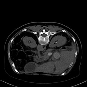
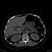
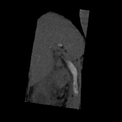
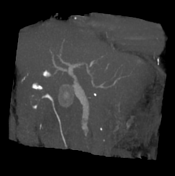

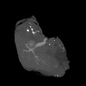
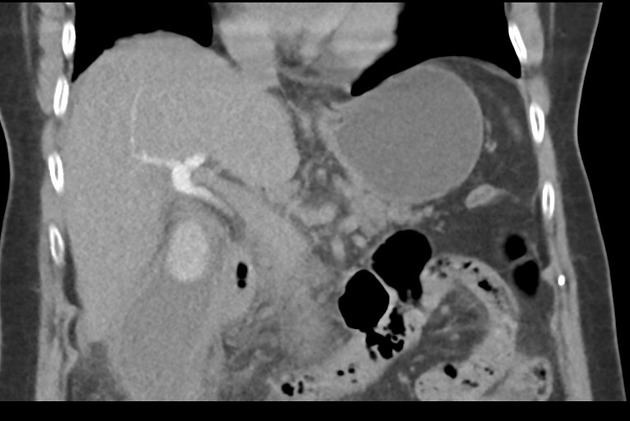
The gallbladder is distended, showing a 2.6 cm calcified stone within, thick walls, and surrounding fat-stranding and free fluid. The common biliary duct is enlarged in caliber (11mm) and demonstrates ill defined filling defects within its distal portions (measuring up to 8 mm on axial plane).
Within the limitations of a non-contrast study the remainder of the imaged upper abdominal content appears unremarkable.
Conclusion: Choledocholithiasis associated with cholecystitis.
Case Discussion
This case illustrates how the CT cholangiography can be useful in identifying choledocholithiasis. This patient has typical CT findings of cholecystitis: thick-walled and distended gallbladder with stones within, as well as surrounding inflammatory fat stranding.


 Unable to process the form. Check for errors and try again.