
Presentation
Transferred from peripheral hospital for investigation of left C2-T1 sensory changes.
Patient Data
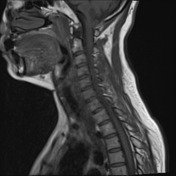


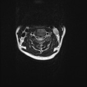
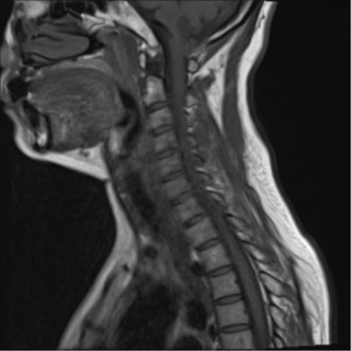
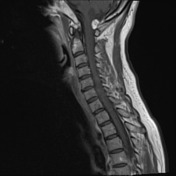
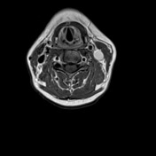
Cervical cord is diffusely expanded to the level of C7-T1 with high T2 signal centrally. Small syrinx present, which only extends to C3-4. A cystic/solid mass is present at the craniocervical junction.
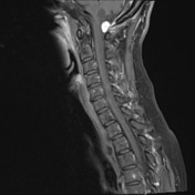
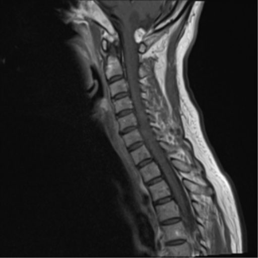
Post contrast images demonstrate the mass has an enhaning nodule and a non-enhancing cystic component.
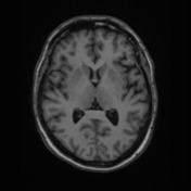
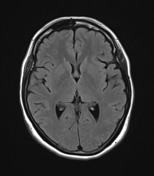

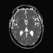
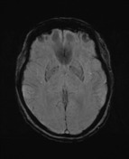
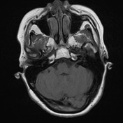

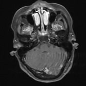
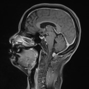
Cystic/solid mass located posteriorly at the craniocervical junction. Larger non-enhancing cystic component which is present anterosuperiorly with a small and intensely enhancing nodule located posteroinferiorly. Cystic component appears intra-axial and separate from the fourth ventricle. The location of the solid nodule is less clear and may be intra-axial with a large eccentric component, or alternatively extra-axial in location posterior to the medulla. No prominent T2 flow voids, susceptibility artefact or diffusion restriction. Medulla is compressed anteriorly with effacement of the premedullary cistern. Abnormal T2 signal throughout the medulla extending into the pons and middle cerebellar peduncles bilaterally.
Histopathology
MACROSCOPIC DESCRIPTION: "Brain tumor": Dusky purple nodule 14x10x7mm with hemorrhagic homogeneous cut surface.
MICROSCOPIC DESCRIPTION: The sections show a well-demarcated nodular tumor with scattered stromal cells in a background of many capillary-sized blood vessels. The tumor cells have mildly enlarged nuclei with hyperchromasia and foamy vacuolated cytoplasm. Mitoses are inconspicuous. There is no necrosis. The features are those of hemangioblastoma. There is no evidence of malignancy. Immunostains are to follow.
DIAGNOSIS: Brain tumor: Hemangioblastoma (WHO Grade I).
Case Discussion
Although the location is unusual (and probably arises from the medulla), the imaging features are fairly typical for hemangioblastoma, with the only characteristic feature lacking being prominent T2 flow voids.


 Unable to process the form. Check for errors and try again.