
Presentation
Fall from 3 meters high.
Patient Data
Age: 85 years
Gender: Male
From the case:
Extrapleural hematoma

Download
Info
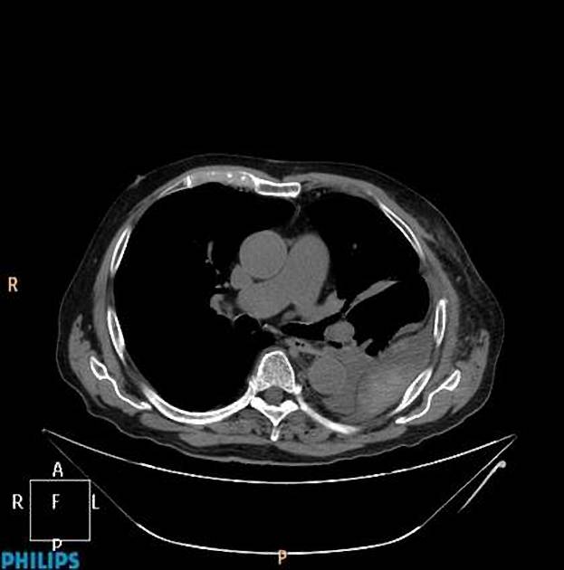
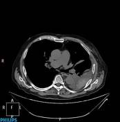
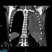
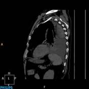
Non contrast CT axial, coronal and sagittal images show biconvex hematoma, which displaces the parietal pleural and the extrapleural fat.
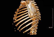
In addition multiple rib fractures are seen.
Case Discussion
Extrapleural hematoma is usually caused by trauma. It is produced by rupture of intercostal or internal mammary vessels. There is no interruption of the parietal pleura, which allows blood to stay in the extrapleural space. It is usually associated with rib fractures.
The observed very thin hypodense line representing the parietal pleura is a helpful imaging feature of these extrapleural collections.


 Unable to process the form. Check for errors and try again.