
Presentation
Severe bilateral flank pain, worse on movement. No previous spinal surgery, neurology or urinary symptoms. Previously fit and healthy.
Patient Data


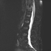
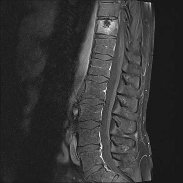
Abnormal enhancement and marrow signal in the T11 vertebral body with intact endplates. Calcific densities within the disc with herniation into the superior endplate of the T11 vertebra.
Given the clinical history, the findings are felt to represent a Schmorl node.
Case Discussion
Clinical correlation:
The patient had unremarkable blood tests including a negative TB quantiferon assay. Normal CT abdomen / CT IVP and normal colonoscopy.
Repeat imaging and follow up:
There was an interval decrease in edema and enhancement within the T11 vertebral body between the initial MRI scan and one performed 3 months later. No other new abnormalities were demonstrated. This strengthens the favored diagnosis of resolving inflammatory changes of a Schmorl node.


 Unable to process the form. Check for errors and try again.