
Presentation
The patient is smoker presented with neck mass and history of right ear pain.
Patient Data


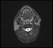
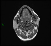
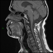
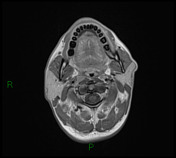
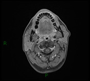
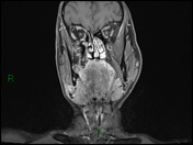
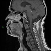
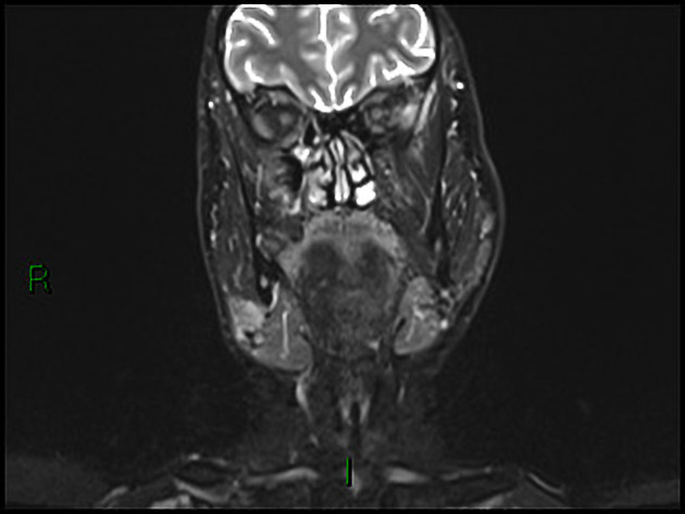
There is abnormal signal intensity mass seen involving the right aspect of the nasopharynx centered on the right fossa of Rosenmuller appears of low signal intensity in T1, intermediate signal intensity in T2 with early post contrast enhancement.
The described mass has the following extensions:
- Anteriorly: extending to the posterior choanal orifice of he nasal cavity and infiltrating the right medial pterygoid plate.
- Posteriorly: the lesion is abutting the prevertebral muscle with no MRI evidence of extension.
- Laterally: the lesion involving the levator palatini muscle and pharyngo-basilar fascia.
- Superiorly: no MRI evidence of intracranial extension.
- Inferiorly it is extending to the upper oropharyngeal mucosa.
- Posterolaterally the lesion involving the petrous apex abutting the medial aspect of the internal carotid artery at its petrous course.secondary right mastoiditis is also noted. Altered marrow signal is also noted within the clivus likely due to marrow infiltration by the tumor.
- No extension across the midline noted.
Bilateral enlarged retropharyngeal lymph nodes the largest measures about 1 x 1.3 cm on the left side. Bilateral enlarged upper and lower deep cervical lymph nodes (bilateral level II, right level IB, V, III and IV). The largest measures about 2 x 2.6 cm on the right side.
Case Discussion
The biopsy of this case showed undifferentiated non keratinizing carcinoma with dense stroma composed of lymphoid cells, which is usually associated with EBV infection. MRI staging is T3 N2 Mx.


 Unable to process the form. Check for errors and try again.