
Presentation
Cough and shortness of breath for month but worse over two weeks. Sudden worsening of breathlessness today, increased oxygen requirements. Left sided chest pain. Hx atrial fibrillation. For CTPA please - high risk PE.
Patient Data
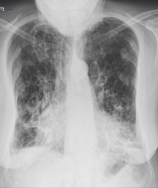
Cardiomegaly with a cardiothoracic ratio of 16:27.
The lungs are hyperinflated and show extensive bilateral ring shadows consistent with bronchiectasis. Bibasal linear opacities are in keeping with atelectasis. Upper zone scarring is likely a result of previous TB.
Bilateral old rib fractures.


No pulmonary thromboembolism.
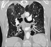
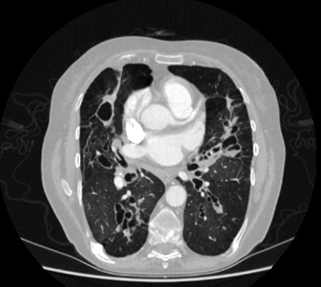
There is severe cystic bronchiectasis, most prominent within the right middle lobe and in the lower lobes bilaterally. There is some mucus plugging within some of the bronchiectatic bronchi in the lower lobe lobes.
There is some apical fibrous scarring related to the prior TB. Multiple areas of scarring are also noted at the lung bases bilaterally. There are air-fluid levels in several of the larger bronchi but no evidence of a mycetoma. No suspicious pulmonary mass lesion is identified.
There is a mosaic attenuation pattern with ground-glass opacity predominantly in the right upper lobe. These changes are likely secondary to gas trapping and superimposed infection.
Multiple healed bilateral posterior rib fractures and there is a kyphosis, with mild anterior wedging of several mid thoracic vertebrae. Incidental hepatic cysts.
Opinion:
Severe cystic bronchiectasis. No pulmonary thromboembolism. Ground glass opacity in the right upper lobe is in probably in keeping with acute infection.
Case Discussion
This case demonstrates the hallmarks of cystic bronchiectasis with bronchial wall thickening and dilated "cysts" visible on the chest x-ray, these correspond to fluid filled cystic bronchiectasis on CT.
The three subtypes of bronchiectasis, based on morphology are:
- cylindrical bronchiectasis
- commonest form, bronchi have a uniform caliber, do not taper and have parallel walls (tram track sign and signet ring sign)
-
varicose bronchiectasis
- uncommon, beaded appearances where dilated bronchi have interspersed sites of relative narrowing
- cystic bronchiectasis
- severe form with cyst-like bronchi that extend to the pleural surface
- air-fluid levels are commonly present


 Unable to process the form. Check for errors and try again.