
Presentation
Poor oral intake, refusal to walk and right-sided abdominal pain for 4 days.
Patient Data

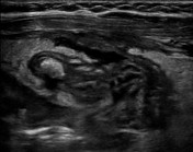
Ultrasound of the right lower quadrant demonstrates an enlarged appendix (anteroposterior diameter > 11 mm) with surrounding periappendiceal hyperechoic structure (PHS) measuring >10 mm.
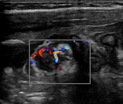
Increased vascularity is seen within the appendiceal wall and the periappendiceal hyperechoic structure (PHS).
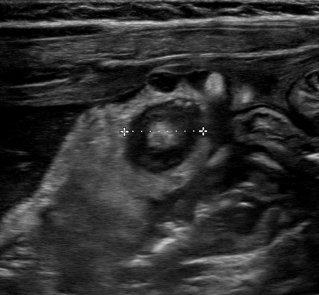
A shadowing fecolith is seen distally within the appendix.
Hypoechoic collections with internal echoes are visualized adjacent to the appendix and PHS.
Case Discussion
The patient went on to have a laparoscopic appendectomy which revealed a gangrenous, perforated appendix with phlegmon.
Periappendiceal hyperechoic structure (PHS) is an ultrasound finding described as an amorphous hyperechoic structure (usually greater than 10 mm) seen surrounding a noncompressible appendix with a diameter greater than 6mm 1.
PHS is thought to represent an inflammatory process of the adjacent omental or mesenteric fat encapsulating the inflamed appendix. It has been associated with severe appendicitis such as gangrenous and phlegmonous appendicitis 1.
Microscopy of appendix indicated ulceration of the mucosa of the appendix and a florid, transmural acute, suppurative inflammatory cell reaction with areas of necrosis of the wall of the appendix and obvious perforation. The inflammation extends into the mesoappendix where there is purulent exudate on the serosal surface, indicating at least localized peritonitis.


 Unable to process the form. Check for errors and try again.