
Presentation
Headaches for long a period.
Patient Data
Age: 20 years
Gender: Male
From the case:
Cerebral arteriovenous malformation


Download
Info

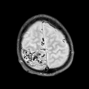
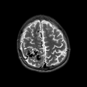
MRI images show a nidus of malformed vessels with the typical appearance of "bag of black worms" in the right parietal region. It originates from the right pericallosal artery and right middle cerebral artery. It drains into superior sagittal sinus. No hemorrhage is seen.
Case Discussion
Typical appearance of cerebral AVM.


 Unable to process the form. Check for errors and try again.