
Presentation
Altered sensation with a vague truncal sensory level.
Patient Data

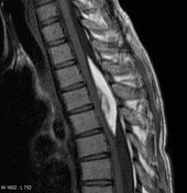
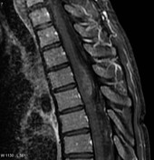
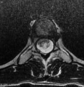
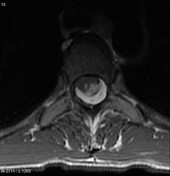
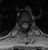
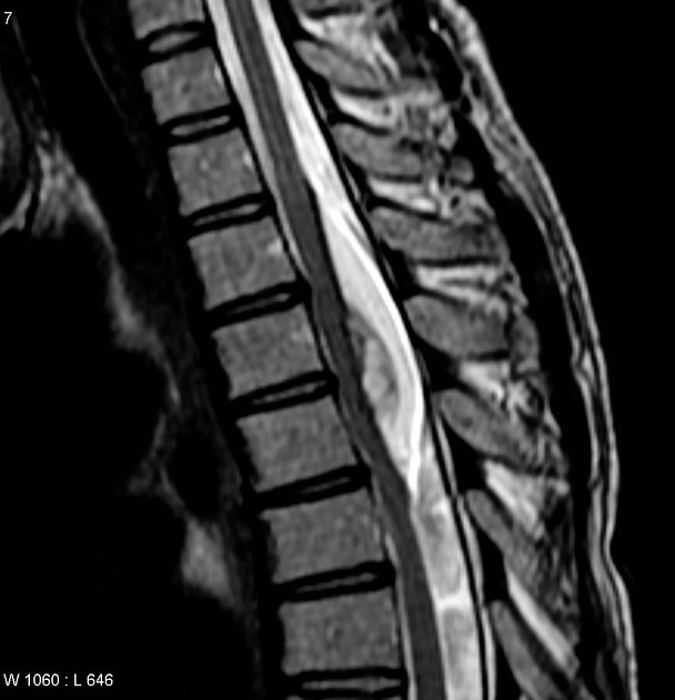
An intradural extramedullary mass having mixed fat and solid enhancing components, displacing and compressing the cord anteriorly.
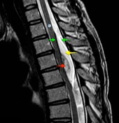
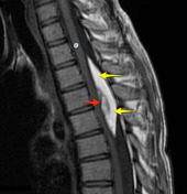
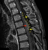
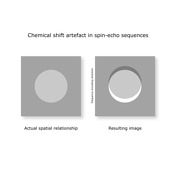

A mass is located behind the spinal cord (C) in an extramedullary intradural location. It is composed of tissue which follows fat on all sequences (bright on both T1 and T2) including fat saturated post contrast T1 (dotted yellow line and arrows). T2 weighted image demonstrates chemical shift artifact around the fatty tissue in the frequency encoding direction (in this diagram anteroposterior). Note how there is signal loss anteriorly and hyperintensity posteriorly (green arrows). This is the result of inaccurate spatial encoding. A smaller anterior component is of intermediate intensity and demonstrated faint contrast enhancement (red arrows).
Case Discussion
There are few fat containing intradural lesions, and the presence of a solid enhancing component excludes a simple lipoma, making the diagnosis almost certainly that of a dermoid, subsequently histologically confirmed.


 Unable to process the form. Check for errors and try again.