
Presentation
Acute abdominal pain in a young man with a known chronic disorder.
Patient Data
Age: 20 years
Gender: Male
From the case:
Sickle cell disease with osseous changes and splenomegaly
Download
Info
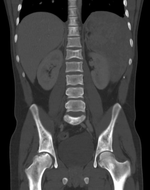
There are FOUR specific findings of sickle cell disease on this single bone window coronal CT:
- splenomegaly - the pain was thought to be due to an acute sequestration crisis
- patchy bone sclerosis
- avascular necrosis of the right femoral head - flattening with subarticular sclerosis and fragmentation
- H-shaped vertebral body - reflecting end-plate micro-infarcts
From the case:
Sickle cell disease with osseous changes and splenomegaly
Download
Info

This annotated image shows the findings of sickle cell disease:
- splenomegaly - the pain was thought to be due to an acute sequestration crisis
- patchy bone sclerosis
- avascular necrosis of the right femoral head - flattening with subarticular sclerosis and fragmentation
- H-shaped vertebral body - reflecting end-plate micro-infarcts
Other findings that may be seen on abdominal imaging (and particularly in radiology exam cases) are gallstones, cholecystectomy clips, and splenic atrophy.
Case Discussion
Chest and abdominal images of sickle cell disease are a real radiology exam favorite - there are often multiple findings which add up to the overall diagnosis.


 Unable to process the form. Check for errors and try again.