
Presentation
Middle aged man with 17 year history of epilepsy. Previous CT 10 years ago apparently normal.
Patient Data
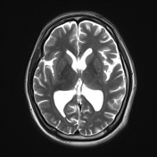

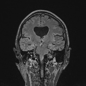

Moderate degree of right perisylvian polymicrogyria. No schziencephaly.
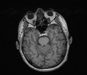
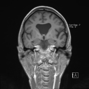
Absent septum pellucidum giving a 'mono-ventricle' appearance, with a normal appearing optic chiasm and no hypotelorism.
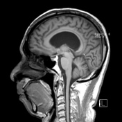
Left hemisphere normal. Posterior fossa normal.
Case Discussion
MRI is now almost routinely performed in those with epilepsy to review for an underlying structural abnormality responsible for seizures.
One of the key elements to be reviewed is the presence of a wide range of cortical dysplasias and migrational disorders. In this case polymicrogyria is found in the most commonly affected area, the perisylvian region, and is associated with an absent septum pellucidum, which is a recognized association.
This is an exceptionally late presentation on MRI, which is likely related to local circumstances and limited access to MRI.


 Unable to process the form. Check for errors and try again.