
Presentation
Community acquired pneumonia. Fever. Rapidly worsening respiratory status.
Patient Data
Age: 21
Gender: Female
From the case:
Pleural empyema
Download
Info
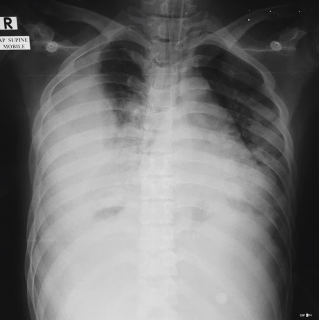
ET and NG tubes.
Bilateral lower lobe consolidation.
Unilateral lenticular shaped right sided loculated pleural collection forming an obtuse angle with the chest wall.
Case Discussion
Pleural empyemas frequently occur in the context of a severe pneumonia.
They tend form an obtuse angle with the chest wall, and due to their lenticular shape and appear loculated along the lateral chest wall.
Ultrasound guided chest drain insertion if often performed in these cases.


 Unable to process the form. Check for errors and try again.