Presentation
Incidentally discovered right adnexal mass on routine ultrasound.
Patient Data
Age: 45 years
Gender: Female
From the case:
Ovarian fibroma
![](https://prod-images-static.radiopaedia.org/images/25561769/ea5408e89a4de9ef8a121f126f064f_thumb.jpeg)
![This study is a stack](/packs/stack-YQKLCKBI.gif)
![](https://prod-images-static.radiopaedia.org/images/25561794/eae617b7de9eac2996b43f45d0335a_thumb.jpeg)
![This study is a stack](/packs/stack-YQKLCKBI.gif)
![](https://prod-images-static.radiopaedia.org/images/25562050/a303f88aa7b3154176898382c1da70_thumb.jpeg)
![This study is a stack](/packs/stack-YQKLCKBI.gif)
![](https://prod-images-static.radiopaedia.org/images/25562415/d5f529b8784ee6a533c8b3c17cc0ec_thumb.jpeg)
![This study is a stack](/packs/stack-YQKLCKBI.gif)
![](https://prod-images-static.radiopaedia.org/images/25562615/c79e33c67e1dbe76663fca6329151a_thumb.jpeg)
![This study is a stack](/packs/stack-YQKLCKBI.gif)
![](https://prod-images-static.radiopaedia.org/images/25562687/36db7d892a83cf7ed3be369105a714_thumb.jpeg)
![This study is a stack](/packs/stack-YQKLCKBI.gif)
![](https://prod-images-static.radiopaedia.org/images/25562747/f5968904a660f813874c6974cbdb86_thumb.jpeg)
![This study is a stack](/packs/stack-YQKLCKBI.gif)
Download
Info
![](https://prod-images-static.radiopaedia.org/images/25561769/ea5408e89a4de9ef8a121f126f064f_big_gallery.jpeg)
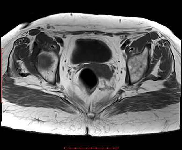
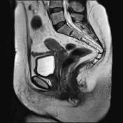
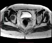
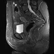
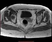
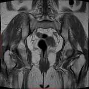
There is a well-defined right adnexal soft tissue mass, eliciting homogeneous low T1 signal intensity, intermediate signal on T2 sequences, with faint post contrast enhancement.
No ascites or pathologically enlarged pelvic lymph nodes.
Case Discussion
A well-defined soft tissue mass with characteristic low T1 and T2 signal intensity is compatible with an ovarian fibrous lesion, such as a fibroma.