
Presentation
Headaches. Prior sinus surgery.
Patient Data


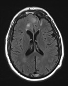
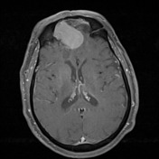
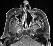
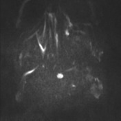
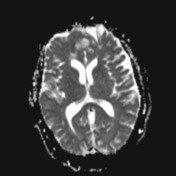
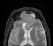
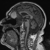
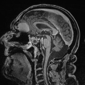
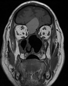
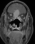
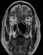
Evidence of frontal and extensive sinus surgery is noted. The right frontal mass, hyperintense on T1, isointense on T2 and post contrast demonstrating no substantial enhancement. A small cystic focus noted posterior to the lesion on T2 imaging. On the left, there is evidence of a prior craniotomy. The frontal pole is distorted and gliotic herniating through a defect in the posterior wall of the frontal sinus. The remainder of the sinus demonstrates high T1 signal consistent with fat.
Findings are consistent with a right sided mucocoele and a left sided encephalocoele.
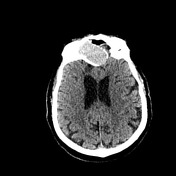
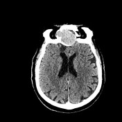

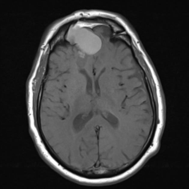
Non-enhancing frontal mass is noted, with significant mass effect on the inferior frontal lobe but no vasogenic edema. Normal venous sinus enhancement is noted. No leptomeningeal enhancement. The midline structures are unremarkable. No extra-axial collection. Previous craniotomy and extensive sinus surgery is noted.
Case Discussion
The patient went on to have surgery which confirmed the presence of a mucocele (which was drained) and defect in the posterior wall of the frontal sinus and encephalocoele. This was repaired. No tumor identified.
Histology
MICROSCOPIC DESCRIPTION: Sections show fibroadipose tissue composed of mature adipocytes and fragments of calcified bone. There is no evidence of atypia or malignancy.
FINAL DIAGNOSIS: Unremarkable fibroadipose tissue, consistent with fat graft.


 Unable to process the form. Check for errors and try again.