
Presentation
History withheld.
Patient Data
Age: 1 year
Gender: Female
From the case:
Retinopathy of prematurity


Download
Info
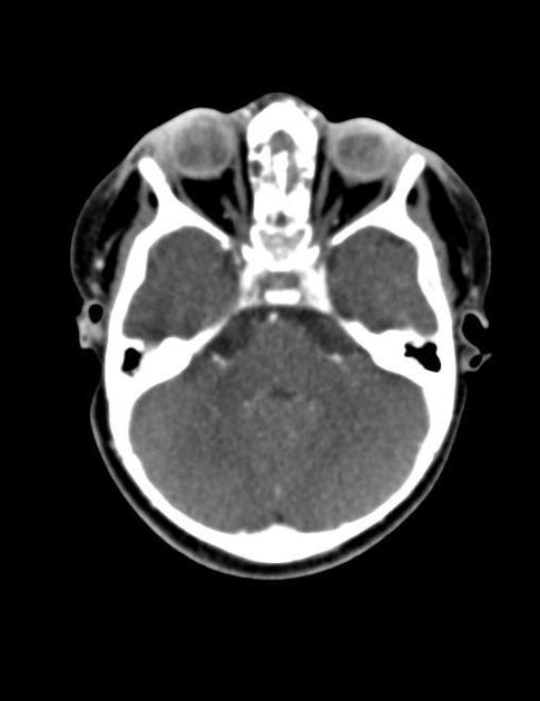
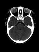
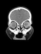
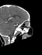
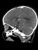
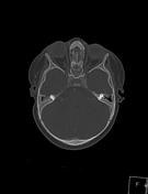
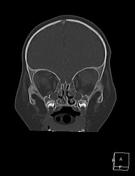
Both eye globes show intra-ocular retro-lental irregular abnormal increased density.
Asymmetrically small left globe.
No evidence of intra-ocular calcifications (which excludes retinoblastoma).
NB:
- bilateral maxillary and ethmoidal sinusitis.
- enlarged nasopahyrngeal adenoids.
Case Discussion
This infant patient was born prematurely and received supplemental oxygen therapy after birth which had led to the development of retinopathy of prematurity.
The retrolental opacity is secondary to prior retinal detachment and fibrovascular organization of the vitreous.


 Unable to process the form. Check for errors and try again.