
Presentation
Short history of abdominal pain. Retrocecal appendicitis?
Patient Data


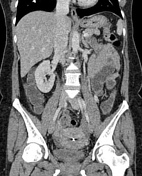
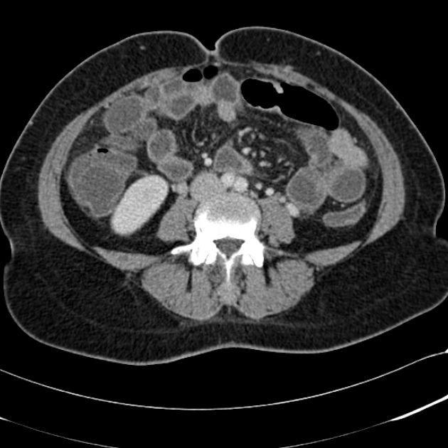
Long thickened inflamed appendix extending medially with the tip lying anterior to the uterus, just superior to the dome of the bladder.
Several appendicoliths within, including an obstructing calcified one in the ostium.
Trace of pelvic free fluid.
No free gas. No collection.
Simple cysts in the liver. The remainder of the solid organs are normal.
IUCD in the uterus.
Case Discussion
CT should be used selectively in acute appendicitis queries.
Clinical examination and ultrasound (especially in the young and thin) being the mainstay.
This is a lovely example of:
- classical signs of appendicitis: thickening appendix, periappendiceal inflammatory change and obstructing appendicoliths
- appendicitis in an atypical location (very anteromedial)
- a very long appendix


 Unable to process the form. Check for errors and try again.