
Presentation
CT colon for investigation of iron deficiency anemia
Patient Data


There is no colonic polyp or mass.
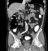
Incidental finding of duplicate IVC. The left common iliac vein continues as the left IVC, receives the left renal vein and then drains into the inferior vena cava. There is no associated abnormality of the kidneys.
Case Discussion
It is important to recognize and report a duplicated IVC as it has implications for future vascular interventions. For example, were IVC filters required, two would need to be placed.
In this particular case, there is no associated renal abnormality such as horseshoe kidney.
This case also illustrates the use of fecal tagging in CT colonography. Gastrografin is used due its tagging and mild laxative qualities. True lesions are differentiated from feces as they won't be tagged and will appear homogeneously of soft tissue density.


 Unable to process the form. Check for errors and try again.