
Presentation
Adult with recurrent chest infections. No history of childhood illness provided. Bronchiectasis?
Patient Data
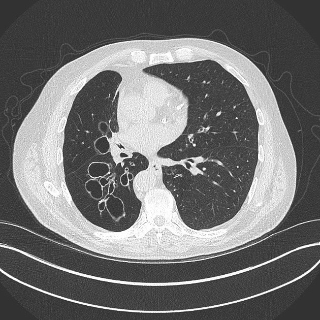
Clusters of cystic spaces of variable size in all lobes of the right lung, most pronounced in the basal segments of the lower lobe. These are thin walled and largely distributed around the distal bronchioles.
The right lung is reduced in volume with smaller and reduced vascular markings with peripheral pruning. This gives a radiolucent appearance to the right lung.
Compensatory hyperinflation of the left lung.
Hiatus hernia.
Unremarkable mediastinum.
Bronchial wall thickened and bronchoceles in the basal segments.
Case Discussion
This is adult presentation of pronounced cystic bronchiectasis.
This patient is likely to have had a childhood LRTI infection resulting in maldevelopment as evidenced by the hyperlucent right lung and cystic changes to the bronchi/oles.
These patients typically get recurrent chest infections.


 Unable to process the form. Check for errors and try again.