
Presentation
CT of the abdomen was performed due to severe epigastric pain.
Patient Data
Age: 55 years
Gender: Male


Download
Info
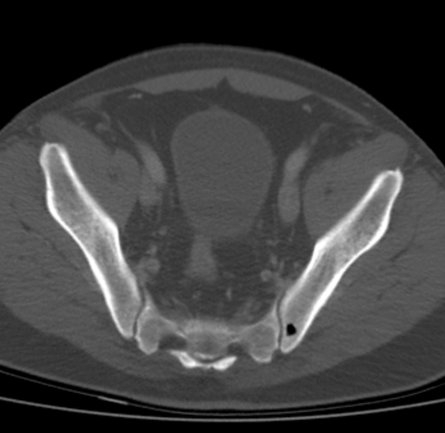
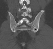
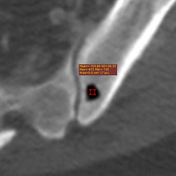
A gas density well-circumscribed lesion is seen in the corpus ossis ilium on the left side. There neither is a surrounding tissue reaction nor a connection to the adjacent sacroiliac joint.
Hounsfield unit value -730 to -672 correspond to gas content.
Case Discussion
Incidental finding of an intraosseous pneumatocyst of the left ilium.


 Unable to process the form. Check for errors and try again.