
Presentation
Unfortunately no clinical history is available.
Patient Data


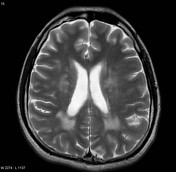
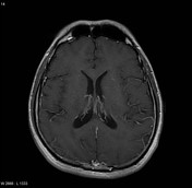
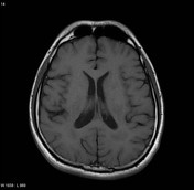
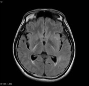

MRI of the brain demonstrates extensive patchy T2 bright white matter change, with involvement also of the posterior limb of the internal capsule. No enhancement.
Case Discussion
Unfortunately I have lost the clinical details of this patient, and all I have available is the results of a dural and brain biopsy, which is non-specific, suggests possible vasculitis, and importantly does not demonstrate any evidence of demyelination, which on imaging would certainly be a possibility.
Histology
These biopsies are indicative of the inflammatory process, of chronic duration, that is not associated with an identifiable microbiologic agent such as acid-fast bacilli, fungi, or toxoplasmosis. The pattern of the inflammatory infiltrate that focally involves vessel walls may be a part of the spectrum of a vasculitic process, either primary or secondary. There was no evidence of a demyelinating disease process. Please correlate these findings with the results of microbiologic studies
Final Diagnosis
- dura: no pathologic diagnosis; crushed (?lymphoid cells)
- brain, leptomeninges and cortex: chronic leptomeningitis and possible vasculitis
- brain, superficial and deep white matter: chronic encephalomyelitis


 Unable to process the form. Check for errors and try again.