
Note: This case has been tagged as "legacy" as it no longer meets image preparation and/or other case publication guidelines.

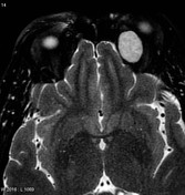

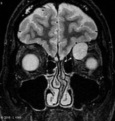
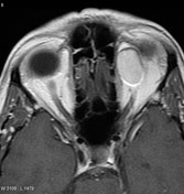
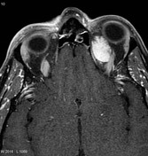
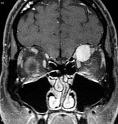
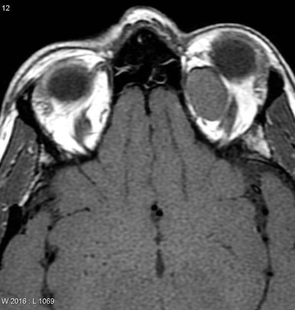
MRI of the orbits with contrast demonstrates a well-circumscribed rounded mass located in the superior medial quadrant in the extraconal space. The mass is intermediate in T1 signal and high signal on T2 weighted images. Contrast-enhanced sequences demonstrate gradual and complete enhancement.
The last obtained sequence is the non-fat suppressed axial T1 image (image 4) also demonstrates a lovely chemical shift artifact at the edges between the mass and the orbital fat.
Features are characteristic of an orbital hemangioma.
Case Discussion
Features are characteristic of an orbital hemangioma. The mass was excised and diagnosis confirmed on histology.


 Unable to process the form. Check for errors and try again.