
Presentation
Intravenous drug abuser, heavy smoker. Fever, purulent infection near right calcaneal tendon. Chest radiograph ordered. Of note, normal radiograph 1 month earlier.
Patient Data
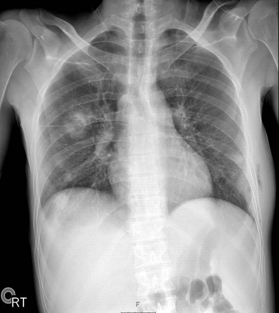
Several round foci in right lung mid- and lower zones and in left lung lower zone. Largest lesion in right lung mid-zone measures 3.7 cm in diameter and clearly cavitary. Lesion projected onto right hemidiaphragm appears cavitary as well.
Lateral chest wall on left is thickened/bulging, with an extracostal vertical line of gas.


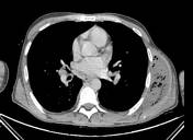
PICC line in superior vena cava.
Multiple well-defined lesions in both lungs, most of which are cavitary and contain fluid levels. Considering patient's history and symptoms and lack of findings on a recent radiograph, most consistent with septic emboli.
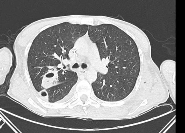
Basal consolidation in right lower lobe (RLL) with subjacent small subpulmonic effusion and adjacent tree-in-bud opacities. Pathogen is most probably mycobacterial or fungal.
Bilateral hilar lymphadenopathy.
Large abscess in lateral left chest wall containing numerous small gas bubbles and tracking paraspinally into posterior left neck (superior aspect falls outside scan) where a gas-fluid level is seen.
Left 5th rib fracture, anterior; 6th rib fracture, anterolateral.
Several tiny gas bubbles in left sternocleidomastoid muscle, perhaps iatrogenic.
Enlarged spleen.


Full resolution of both the lung opacities and the chest wall abscess.
Case Discussion
Due to difficulty in finding a vein, a PICC line was inserted under ultrasound guidance and advanced under angiography (PICC line seen on CT).
The abscess was drained with bacilli cultured from the pus, and the patient went on to receive IV ampicillin and cloxacillin for 3 weeks through the PICC line.


 Unable to process the form. Check for errors and try again.