Presentation
Teenager in RTA with flipped car. GCS low on arrival.
Patient Data
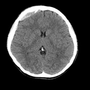

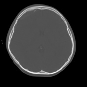

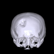
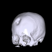
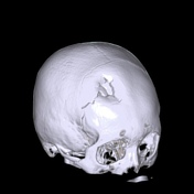

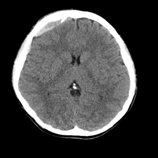
Communiuted depressed right frontal bone skull fracture.
Thin associated subdural hematoma with a small amount of fat layering inside the subdural collection. No significant mass effect.
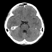
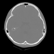
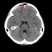
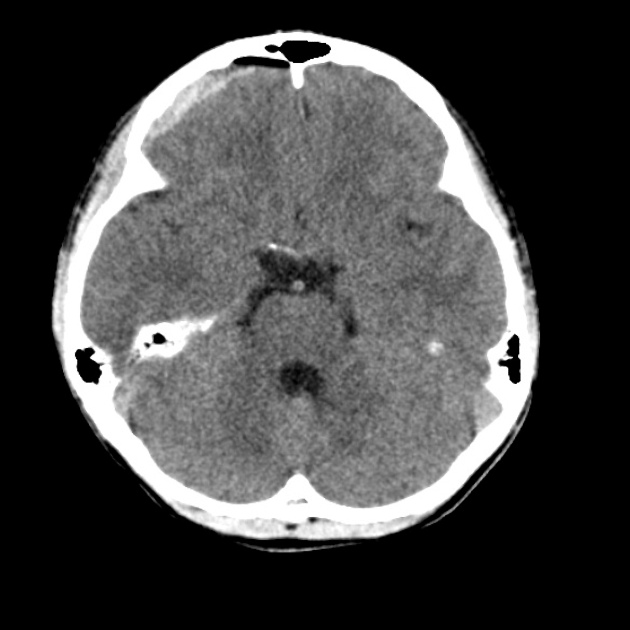
Subdural lipohaematoma: with manual windowing
An interesting rare observation is made of the subdural content.
Initial brain windows suggest a small amount of subdural air, perhaps due to associated fractures of a paranasal sinus.
But on manual windowing this is infact fat layered anteriorly in the subdural hematoma.
The HU using the station 'pixel lens' confirmed this as fat.
Case Discussion
The 3D volume rendered images elegantly illustrate this depressed skull fracture.
The presence of fat inside the subdural hematoma (a subdural lipohaematoma) is an interesting observation as well as a teaching point on the use of CT windows.
The potential mechanism for fat to be present within the subdural is marrow fat or invagination of superficial fat from the depressed fracture.


 Unable to process the form. Check for errors and try again.