
Presentation
Bilateral painful focal foot swelling
Patient Data
Age: 55 years
Gender: Male
From the case:
Tophaceous gout


Show annotations
Download
Info

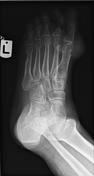
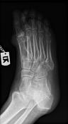
Several well-circumscribed punched-out bony erosions with sclerotic margins exist bilaterally, particularly in the vicinity of the first MTP joints.
Juxta-articular soft tissue densities and swelling are also visible.
Case Discussion
Findings of x-rays are characteristic of tophaceous gout.
The typical appearance of gout on x-ray evaluation is the presence of well-defined, punched-out erosions with sclerotic margins in a marginal and juxta-articular distribution with overhanging edges. Tophi are also pathognomonic features of gout.


 Unable to process the form. Check for errors and try again.