
Presentation
Underlying hypertension. History of sputum negative pulmonary TB and completed treatment. Presented with weight loss and reduced appetite for 1 month. Associated with intermittent fever and hemoptysis. The patient also experienced chest pain.
Patient Data
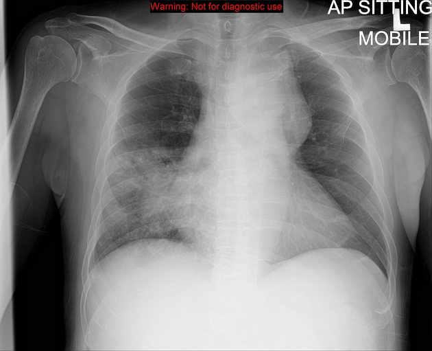
Chest radiograph showed widening of the superior mediastinum with poor visualization of the aortic arch outline suggestive of aortic arch aneurysm. No evidence of hemothorax to suggest ruptured aneurysm.
Ill defined cavity containing soft tissue opacity with surrounding consolidation seen at the right middle zone. No hilar or mediastinal lymphadenopathy.


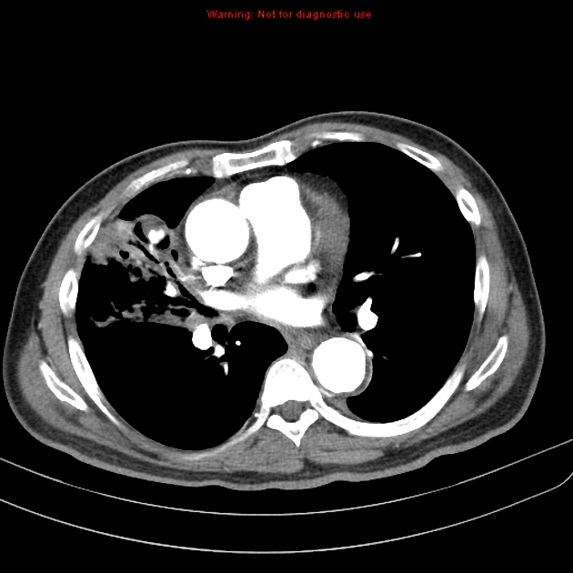
There is a large saccular aneurysmal dilatation of the aortic arch measuring 5.5 (AP) x 5.3 (ML) x 8.4 (CC) cm. Presence of intramural hematoma mainly at the superior aspect. It displaces the trachea to the right side. The aneurysm is in close proximity with the esophagus but no clear communication seen between these structures. No blood products in the esophagus. No contrast extravasation or hemothorax to suggest ruptured aneurysm. Left common carotid and subclavian arteries are normal.
A cavity with surrounding consolidation seen in the right middle lobe. Aneurysmal branch arising from the right middle lobe pulmonary artery seen within the cavity suggestive of Rasmussen aneurysm. Hematoma seen surrounding the aneurysm.
Tree-in-bud appearance and numerous tiny nodules seen in the right middle and lower lobes indicating active endobronchial infection.
Case Discussion
Rasmussen aneurysm is an uncommon complication of pulmonary tuberculosis and represents a pulmonary artery aneurysm adjacent to or within a tuberculous cavity. Usually distributed peripherally and beyond the branches of the main pulmonary arteries.
A weakening of the pulmonary artery wall from adjacent cavitating tuberculosis is the cause of this condition. There is a progressive weakening of the arterial wall as granulation tissue replaces both the adventitia and the media. This is then gradually replaced by fibrin, resulting in thinning of the arterial wall, pseudoaneurysm formation, and subsequent rupture with hemorrhage.
Hemoptysis is the usual presenting symptom and may be life-threatening when it is massive.


 Unable to process the form. Check for errors and try again.