Presentation
Came to ultrasound clinic for follow up. He had surgery to remove an unspecified cyst from the epigastric area 5 years ago. Patient was asymptomatic at the time of ultrasound scan.
Patient Data
![](https://prod-images-static.radiopaedia.org/images/31438037/407e47adf6f9d17f09d70a833faefd_thumb.jpeg)
![](https://prod-images-static.radiopaedia.org/images/31438038/8f8e1cdd20686d5c5bb5fa8b769261_thumb.jpeg)
![](https://prod-images-static.radiopaedia.org/images/31438053/e66ccda7e8b28be0c18b486c48802c_thumb.jpeg)
![](https://prod-images-static.radiopaedia.org/images/31438061/c8deebfb8794bf1f8caa43984a4bbc_thumb.jpeg)
![](https://prod-images-static.radiopaedia.org/images/31438062/05d4fc8ee2f413a546a344d363e16a_thumb.jpeg)
![](https://prod-images-static.radiopaedia.org/images/31438063/127e0224a5f2cdf9f177a8f8915d5f_thumb.jpeg)
![](https://prod-images-static.radiopaedia.org/images/31438085/434853dec829df1387df21fcea7e7b_thumb.jpeg)
![](https://prod-images-static.radiopaedia.org/images/31438093/6daa2b7f47833b53b83303b4534b7b_thumb.jpeg)
![](https://prod-images-static.radiopaedia.org/images/31438125/e0a402e9da80f6cd361762b4b795b0_thumb.jpeg)
![](https://prod-images-static.radiopaedia.org/images/31438094/9db3eb7338d98cf0913e6648ecc58b_thumb.jpeg)
![](https://prod-images-static.radiopaedia.org/images/31438102/33f14c3b49cb33f701604a3839e507_thumb.jpeg)
![](https://prod-images-static.radiopaedia.org/images/31438110/7db3ec552cfd47ac8c826d8cd08545_thumb.jpeg)
![](https://prod-images-static.radiopaedia.org/images/31438133/055e8909aa76ed39817b21f96afb25_thumb.jpeg)
![](https://prod-images-static.radiopaedia.org/images/31438134/d11afd7b9391a5c52366d2f688f192_thumb.jpeg)
![](https://prod-images-static.radiopaedia.org/images/31438135/2d142505d7204c40273b52939d8c3e_thumb.jpeg)
![](https://prod-images-static.radiopaedia.org/images/31438136/c1167eb3c62f3e2a5074f3cdd74723_thumb.jpeg)
![](https://prod-images-static.radiopaedia.org/images/31438037/407e47adf6f9d17f09d70a833faefd_big_gallery.jpeg)
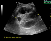
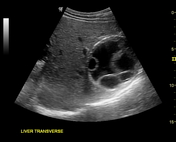
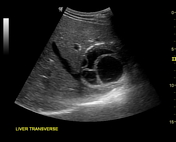
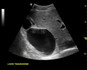
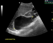
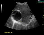
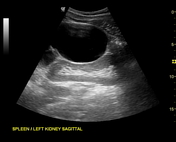
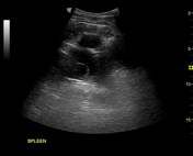
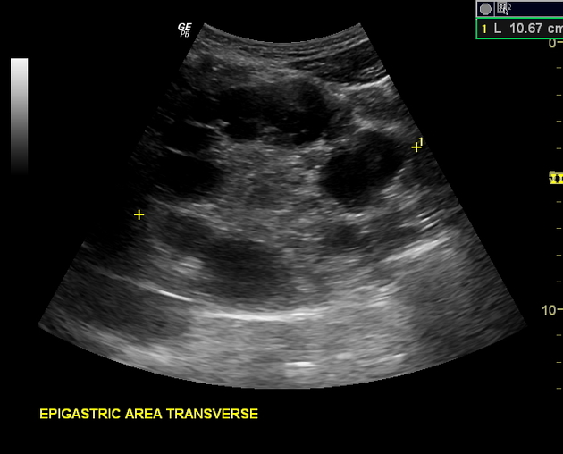
Multiple hydatid cysts of variable sizes, sonographic appearance and stages seen in the liver, spleen, epigastric area (possibly retroperitoneal), pelvic cavity and anterior to the left psoas muscle (possibly retroperitoneal).
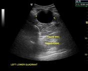
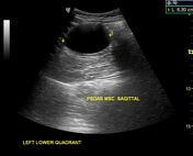
Unilocular anechoic cyst (stage CL) according to WHO classification seen anterior to left psoas muscle at retroperitoneum.
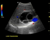
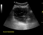
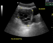
Unilocular mother cyst with honeycomb appearance (stage CE 2) (according to WHO classification) seen in the pelvis, just cranially to the urinary bladder.
Case Discussion
Disseminated hydatid disease, also known as disseminated abdominal hydatidosisis, is a rare presentation of hydatid disease. Secondary peritoneal disease usually occurs as a result of either traumatic or surgical rupture, as in this case, of a primary hepatic or splenic cyst. The appearance of hydatid cysts on ultrasound can be classified according to World Health Organization 2001 classification of hepatic hydatid cysts. In this case, different stages of the WHO classification can be seen.