
Presentation
No history provided.
Patient Data
Age: Young adult
Gender: Female
From the case:
Neurofibromatosis type 1

Download
Info

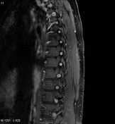
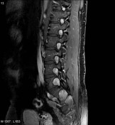
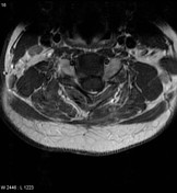
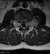
Selected images from an MRI of the spine with contrast demonstrate extremely widespread enhancing masses occupying almost every single neural exit foramen and in some cases resulting in canal stenosis.
Note: This case has been tagged as "legacy" as it no longer meets image preparation and/or other case publication guidelines.
Case Discussion
This patient has a known diagnosis of neurofibromatosis type 1 (NF1). Phakomatoses often result in some of the most impressive scans, and as many patients have recurrent problems from the numerous tumors they develop, they are frequently and serially imaged.


 Unable to process the form. Check for errors and try again.