
Presentation
Bitemporal hemianopia
Patient Data
Age: Adult
This case has been tagged as "legacy" as it no longer meets image preparation and/or other case publication guidelines.

Download
Info
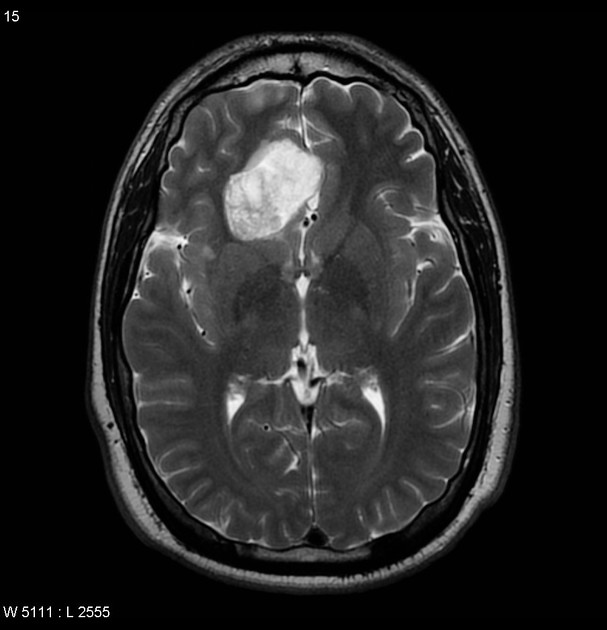
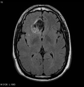
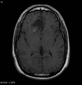
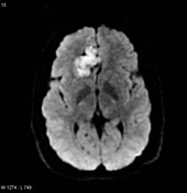
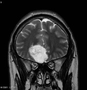
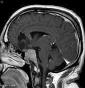
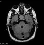
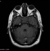
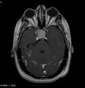
At the medial aspect of the right frontal lobe a lobulated lesion demonstrates predominantly low T1 and very high T2 signal with little if any enhancement. Diffusion weighted imaging reveals prominent restricted diffusion.


Download
Info

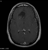
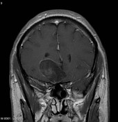
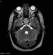
Solid and homogeneously enhancing mass enlarging the pituitary fossa, with suprasellar extension, and partial encasement of the right ICA.
Case Discussion
Appearances of the frontal lobe mass are most consistent with an epidermoid cyst.
Additionally as pituitary mass is noted, consistent with a coincidental pituitary macroadenoma.


 Unable to process the form. Check for errors and try again.