
Patient Data
Age: Adult
- Note: This case has been tagged as "legacy" as it no longer meets image preparation and/or other case publication guidelines.
From the case:
Ganglioglioma

Download
Info
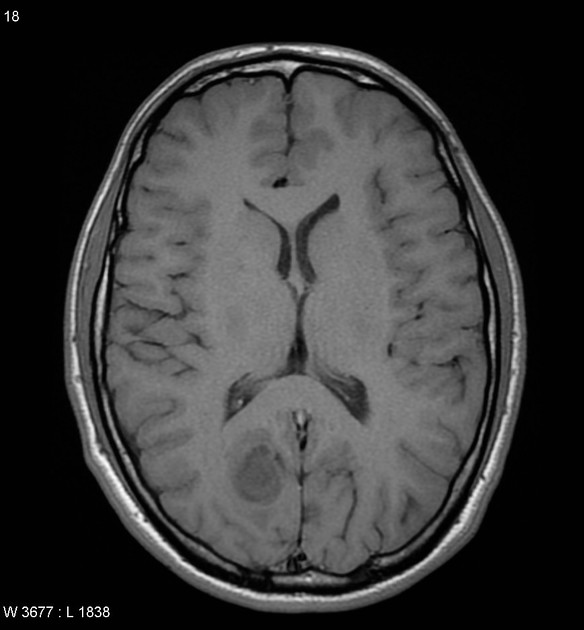
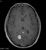
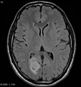
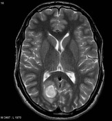
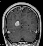
MRI of the brain demonstrates a well circumscribed mass appearing to be located within the cortex (in a sulcus) of the right occipital lobe. It slightly hypointense to adjacent grey matter, hyperintense on T2 weighted images and demonstrates homogeneous contrast enhancement with only as small amount of surrounding edema.
Case Discussion
Appearances suggest a cortical tumor, such as a ganglioglioma (subsequently histologically proven).
Patient went on to have a local recurrence 18 months later which was predominantly glial.


 Unable to process the form. Check for errors and try again.