
Presentation
Abdominal pain. End stage renal disease and hypothyroidism. No alcohol consumption.
Patient Data
Age: 20 years
Gender: Female
From the case:
Chronic pancreatitis

Download
Info
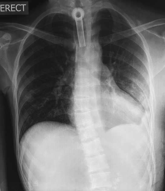
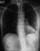
Chest x-ray shows:
- tracheostomy tube
- left basal pleural thickening
- lungs clear
- punctate calcification in the upper abdomen in the midline
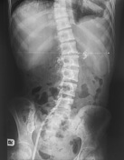
Abdominal x-ray shows right femoral line, in addition, to punctuate calcifications in the midline of the upper abdomen following the morphology of the pancreas.
Download
Info
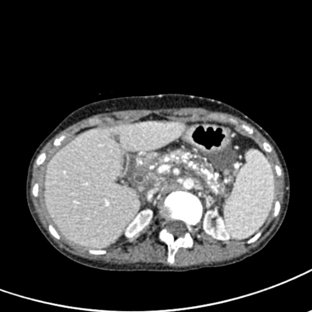
Punctate calcification throughout the pancreas.
No gallstones.
The kidneys are atrophic.
Small amount of abdominal free fluid.
Case Discussion
The appearances are of chronic pancreatitis.
Worldwide, alcohol excess is the commonest cause although there is a wide differential.
Starting with the CXR this makes a decent examination viva case:
- spot the film edge (review area) of the chest radiograph abnormality of upper abdominal calcification
- this earns the abdominal radiograph to permit confirmation
- CT follows to facilitate a discussion on the causes of pancreatic calcification and the causes of chronic pancreatitis


 Unable to process the form. Check for errors and try again.