
Presentation
Imaged for headaches.
Patient Data
Age: 50 years old
Gender: Male
From the case:
Asymmetric fatty marrow in the petrous apex


Download
Info
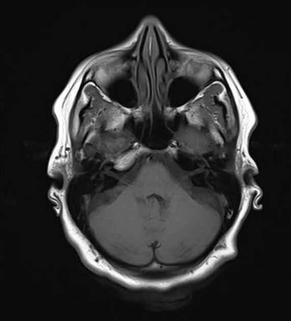
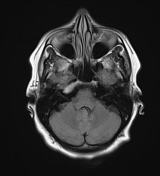
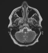
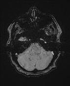
Unenhanced T1 & T2 weighted images reveals smoothly marginated T1w hyperintense signal in right petrous apex (fat signal).
Case Discussion
Asymmetric fatty marrow of right petrous apex.
Asymmetric pneumatization is related to another normal variation: asymmetric fatty marrow within the petrous apex. This finding is a common incidental finding on brain, skull base, and soft-tissue neck MRI studies obtained for evaluation of nonotologic complaints. Typically, normal marrow contains significant adipose tissue, and signal characteristics parallel those of scalp or orbital fat. Fatty marrow is hyperintense on routine T1- and T2-weighted sequences. (1)


 Unable to process the form. Check for errors and try again.