
Presentation
Long history of left loin pain, no history of dyspnea, cough or fever.
Patient Data
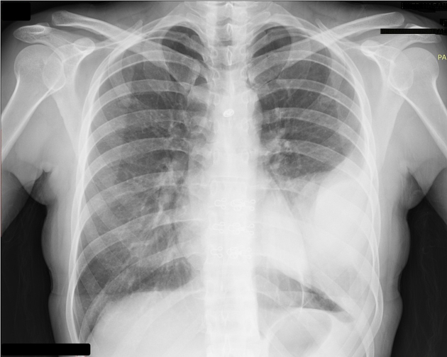
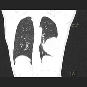
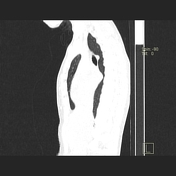
A well-defined round shaped pleural based mass lesion is seen in the left side of the chest, It shows homogeneous opacity with smooth margins and no matrix of calcification detected, the findings are in favor of hydatid cyst (CT chest with contrast was recommended).
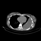


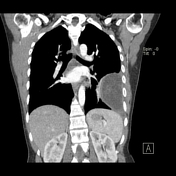
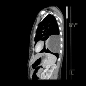

A well-defined oval-shaped large intra-pleural cystic lesion with numerous daughter cysts inside is seen on the left side of the chest. It showed fluid CT density, smooth margin, and hypodense content relative to the capsule with a thin enhancing capsule, picture highly in favor of an uncomplicated pleural hydatid cyst.
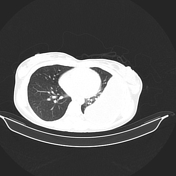
Both lungs are clear with normal pulmonary vascular markings, and no focal lesion.
Normal mediastinum with normal mediastinal vasculature, no mass or enlarged lymph nodes.

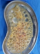
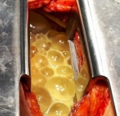
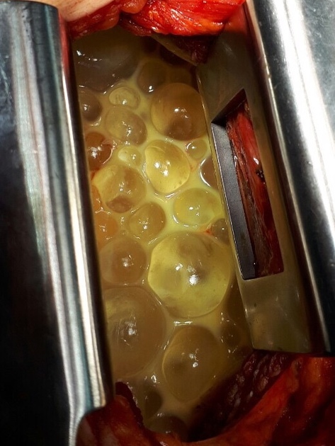
The patient underwent thoracotomy and the findings of numerous cystic lesions within the pleura confirmed the diagnosis of hydatid cysts.
Case presented with permission, courtesy of Dr Chalak (cardiothoracic surgeon).
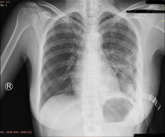
Left lung shows haziness in lower zone with blunting of left costophrenic angle secondary to pleural effusion. Right lung clear and right costophrenic angle within normal limits. Both hilum normal in size and density. Drainage tube is seen in left side of the chest wall.
Case Discussion
Hydatid cysts result from infection by the Echinococcus worm and can result in cyst formation anywhere in the body.
Primary pleural hydatid cyst is an uncommon mode of presentation of hydatid disease.
It is important to keep hydatid disease in mind as one of the rare but possible differential diagnoses in cystic lesions of the chest wall in the endemic zones. Blind aspiration should be avoided.


 Unable to process the form. Check for errors and try again.