
Presentation
Right-sided blurring of vision.
Patient Data
Age: 25 years
Gender: Male
From the case:
Choroidal melanoma


Download
Info

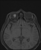
The study shows a well defined intra-ocular T1 hyperintense and T2 hypointense lesion along the superomedial aspect of right eye showing mild enhancement on the postcontrast study.
Case Discussion
Choroidal melanomas, also called malignant uveal melanomas, are the most common primary ocular tumor in adults.
On imaging, melanomas appear hyperdense on CT scan and hyperintense on T1WI on MRI. Mild enhancement can be seen on post-contrast images.
Choroidal melanomas can be associated with retinal detachment on presentation. Systemic metastasis may occur in the liver, lung, bone, kidney and brain.


 Unable to process the form. Check for errors and try again.