
Presentation
CSF rhinorrhea of several months. No history of trauma.
Patient Data
Age: 40 years old
Gender: Male
From the case:
Frontoethmoidal encephalocele


Download
Info
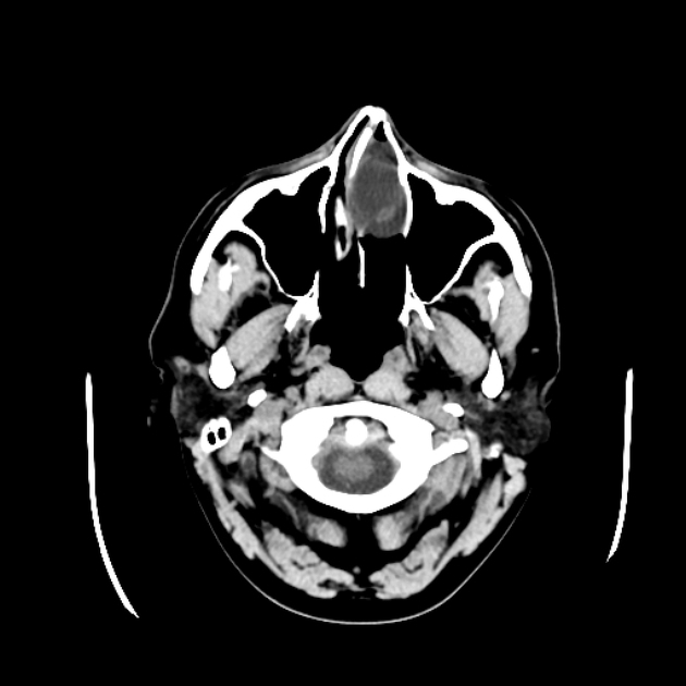
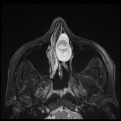
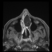
A cystic lesion is seen filling the left nasal cavity.
A bone defect is noted at the left aspect of cribriform plate through which this cystic lesion is arising.
From the case:
Frontoethmoidal encephalocele


Download
Info
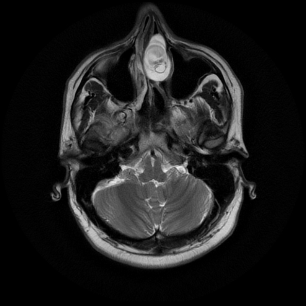
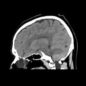
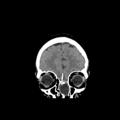
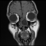
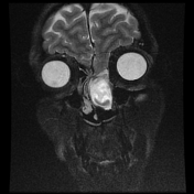
A CSF containing cyst is seen herniating through the cribriform plate into the nasal cavity containing part of the frontal lobe.
Case Discussion
Frontoethmoidal encephalocele is a sac containing CSF, meninges and brain tissue herniating through a bone defect at the nasal or frontal bones. It can occur spontaneously, be post traumatic or secondary to raised intracranial pressure e.g. tumor.


 Unable to process the form. Check for errors and try again.