
Presentation
Increased frequency of frontal tension headaches. Rule out intracranial lesion.
Patient Data
ASNR Case of the Day Winner 2018
This case was selected as the winner of the 2018 ASNR Case of the day competition winner. Runner-up and honorable mentions can be viewed in this playlist.


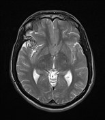
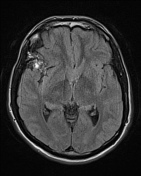

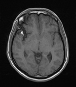
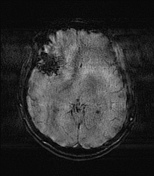
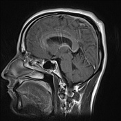
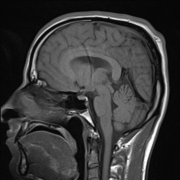
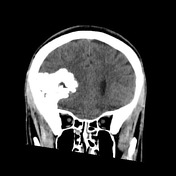
There is a large extra-axial calcified mass overlying the right frontal lobe. It is mostly calcified with expected hypointense signal on T1/T2 within areas of calcifications and diffuse susceptibility artefacts on SWI. It is mildly T1 hypointense, moderately T2 hyperintense (outside of the calcific foci) and demonstrates mild internal enhancement. A small pedicle arising from right greater wing of sphenoid is noted. There is no diffusion restriction.
There is no significant intraparenchymal edema and no other abnormal intracranial enhancement.
There is moderate mass effect on the adjacent right frontal lobe parenchyma with mild rightward midline shift and compression of the right lateral ventricle (especially the frontal horn).

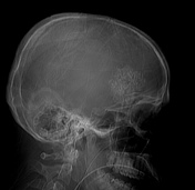
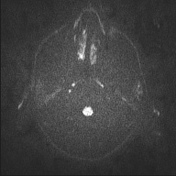
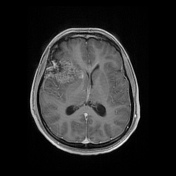
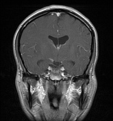
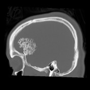
Scout demonstrates extensive calcifications within the large extra-axial lesion. These calcifications have a pattern that suggests a chondroid matrix.

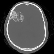
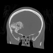

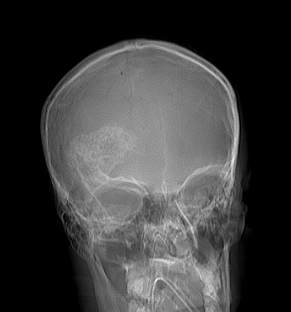
There is a large extra-axial calcified mass overlying the right frontal lobe. The calcifications have a ring and arcs pattern, which suggests a chondroid matrix. There is an osseous pedicle at the inferior aspect of the mass which is continuous with the superomedial aspect of the right sphenoid wing. There is no hyperostosis or remodeling of the cranial vault.
There is moderate mass effect on the adjacent right frontal lobe parenchyma with mild rightward midline shift and compression of the right lateral ventricle (especially the frontal horn).
There is no other intracranial or osseous abnormality.
Case Discussion
The findings suggest a pedunculated chondroid lesion arising from the internal aspect of the right sphenoid wing, most likely chondrosarcoma. The appearance of the calcifications would be very atypical for meningioma and chondromyxoid fibroma which are the main differential diagnoses.
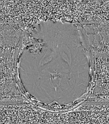
MRI is shown first on purpose as the CT-scan best demonstrates the underlying chondroid matrix ― less definite and much more puzzling on MRI.
Histopathology
Microscopic description:
- lobules of cartilage and some moderately cellular myxoid stroma extensively permeating and disrupting lamellar bone
- lobules of cartilage are of low to moderate cellularity and the chondrocytes show mild and moderate cytologic atypia
- there are foci of old necrosis with infiltration by foamy histiocytes
- no cytologic atypia is seen in the bone
Final diagnosis: Intermediate chondrosarcoma (grade II)


 Unable to process the form. Check for errors and try again.