
Presentation
Headache. History of focal seizures in childhood.
Patient Data
Note: This case has been tagged as "legacy" as it no longer meets image preparation and/or other case publication guidelines.
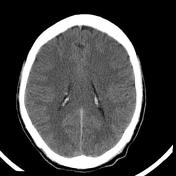

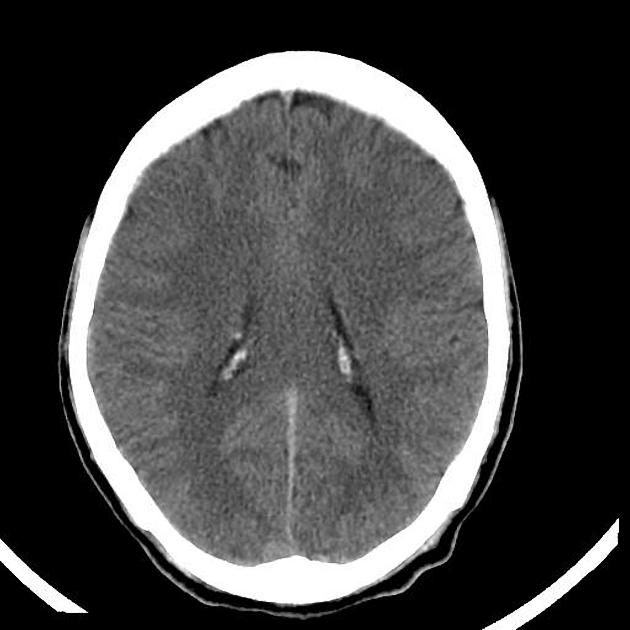
CT reveals a solitary calcified subependymal lesion on body of right lateral ventricle and a hyperdense lesion at the foramen of Monro on left side.
Case Discussion
Patient was being treated for seizure disorder and also had cutaneous manifestations of Tuberous Sclerosis.
There was h/o focal seizures in childhood. The patient also had Shagreen patches and adenoma sebaceum. MRI done subsequently showed additional cortical tubers.
SGCA is the second most common manifestation of tuberous sclerosis (TS). First being tubers. It affects 5% of patients. The foramen of Monroe is the classic location. Some of the subependymal nodules themselves can transform into SGCA over a period of time.


 Unable to process the form. Check for errors and try again.