
Presentation
Orbital mass growing by the time.
Patient Data
Age: 45 years
Gender: Female
From the case:
Orbital rhabdomyosarcoma


Download
Info
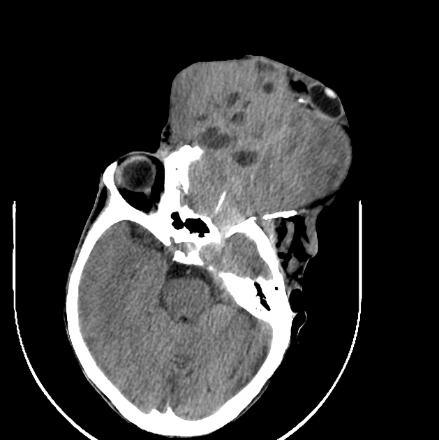
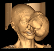
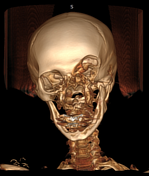
There's a huge mass with necrotic and calcified lesions inside, expanding left orbit, with the displacement of left eye globe anteriorly. The mass is involving the paranasal sinuses and the nasal cavity. Destruction is seen in the frontal and maxillary bones.
Case Discussion
The most probable diagnosis is rhabdomyosarcoma. Neurofibroma is in the differential diagnosis.


 Unable to process the form. Check for errors and try again.