
Presentation
Abdominal pain. History of appendectomy 20 years prior.
Patient Data
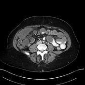

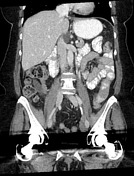
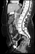

Mildly dilated proximal small bowel with gradual admixture of oral contrast with enteric fluid as the bowel progresses distally. Two adjacent angulated transition points entering and exiting correspond to the "closed loop" segment, and form a "waist"-like appearance in the right mid abdomen on the axial images. The closed loop portion is dilated, fluid-filled, and has mesenteric edema/interloop fluid. No pneumatosis.
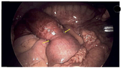
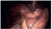

Laparoscopic images from surgery indicated a tight adhesive band as the cause of obstruction (arrows).
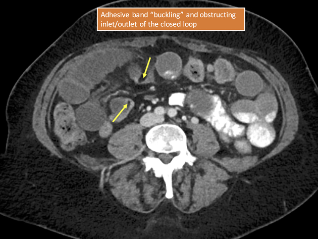
Arrows indicate the site of small bowel entering the internal hernia defect. Note how both segments of bowel are angulated toward each other.
Case Discussion
There are several subtle clues indicating a closed-loop obstruction in this case. The proximal small bowel is mildly dilated, but less so than the closed loop segment in the right lower quadrant. The small bowel in the closed loop sticks out as more abnormal than any other segment in the abdomen: more dilated, no oral contrast, and mesenteric edema. The axial images give it away (see annotated image) - the two sites of obstruction are adjacent to each other, indicating a closed loop obstruction. This appearance could be due to internal hernia or (as in this case) adhesive bands.


 Unable to process the form. Check for errors and try again.