
Presentation
Weight loss work up.
Patient Data


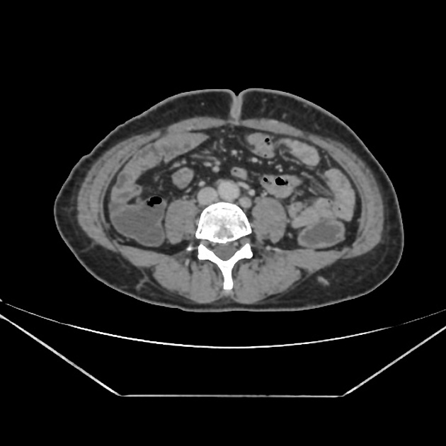
Incidental note is made of duplication of inferior vena cava with right IVC draining right external iliac vein. Left inferior vena cava drains left renal vein, left common iliac vein and right internal iliac vein. Left IVC rejoins right IVC in the infrahepatic portion.
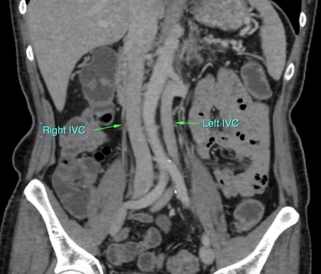
Annotated depiction of three vessel pattern in the abdomen, wherein the abdominal aorta is sandwiched between two IVCs.
Case Discussion
This was an incidental finding picked up in a patient with weight loss work up. A poorly defined lesion was identified in the pancreatic body with a long segment circumferential mural thickening in sigmoid. Few focal liver lesions are present, some typical of hemangiomas, others possible secondaries.
Identification of duplication of IVC warrants further detailed evaluation of abdomen to rule out any other associated urogenital developmental anomalies.


 Unable to process the form. Check for errors and try again.