Presentation
Right-sided hemiparesis, aphasia, and mouth deviation.
Patient Data
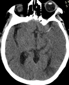

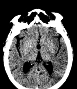

A large area of hypo-attenuation with loss of gray-white matter differentiation involving the left temporal, parietal, and occipital lobes, and the caudate nucleus. These changes are more evident in the stroke window.
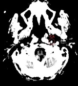
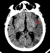
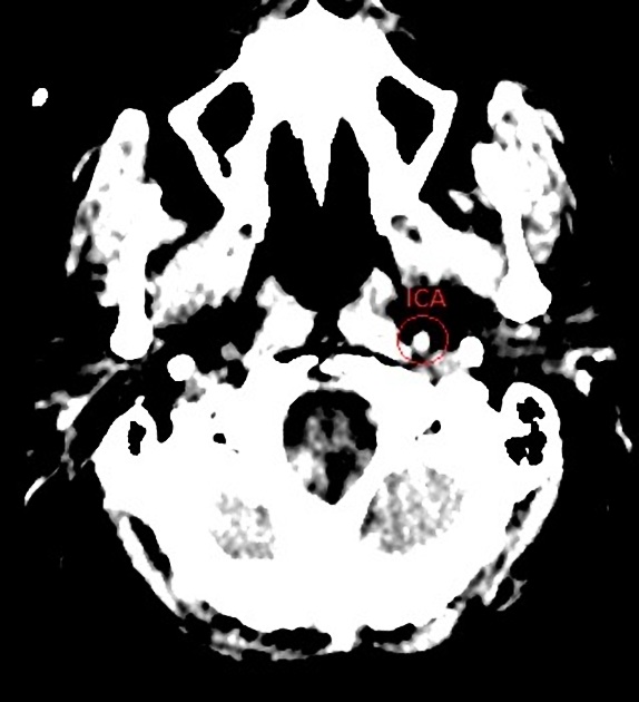
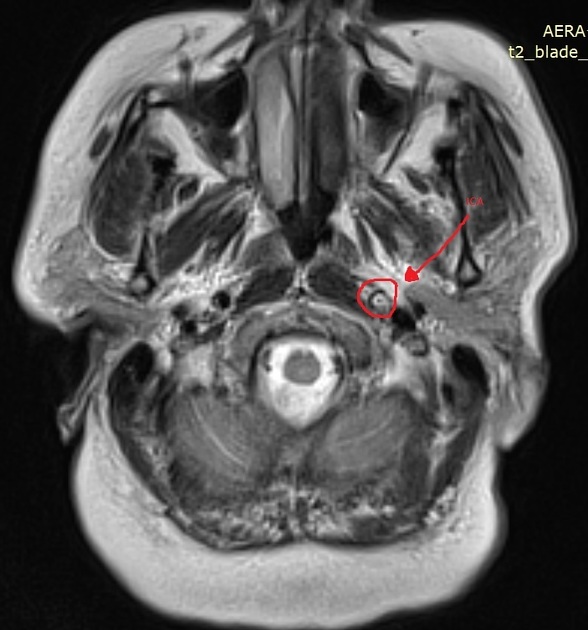
First three annotated images show hyper-attenuation of the left internal carotid artery and left middle cerebral (M1 and M2 segments) artery. This is known as the hyperdense MCA sign.
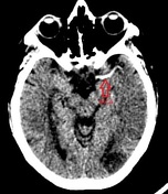
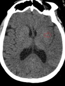
The fourth annotated image shows loss of differentiation of the gray-white interface in the lateral margin of the left insular cortex. This is known as the loss of the insular ribbon sign.

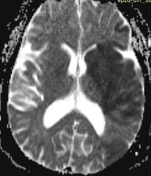

High diffusion signal involving the entire left caudate nucleus and cortex of left temporal, parietal and occipital lobes.

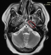
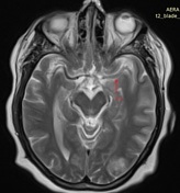
Hyperintensity along the intraluminal part of the left internal carotid artery and left middle cerebral artery.
Case Discussion
Non-enhanced CT scan is the initial step to rule out intracranial hemorrhage during a 'stroke call', and can demonstrate some clear signs of ischemic stroke.
This case illustrates the classic signs of middle cerebral artery territory infarct, such as the loss of the insular ribbon sign, the hyperdense MCA sign, and loss of gray-white matter differentiation. Adjusting the window and level (e.g. W:40 L:40 or W:8 L:32) is used for optimized visualization of subtle loss of gray-white matter differentiation.
Restriction diffusion on DWI and ADC maps are in-keeping with extensive acute ischemic infarction of left middle cerebral artery territory.


 Unable to process the form. Check for errors and try again.