
Presentation
Treated for chest infection. Clinician has concerns on the chest radiograph of a pleural effusion.
Patient Data

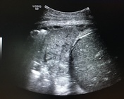
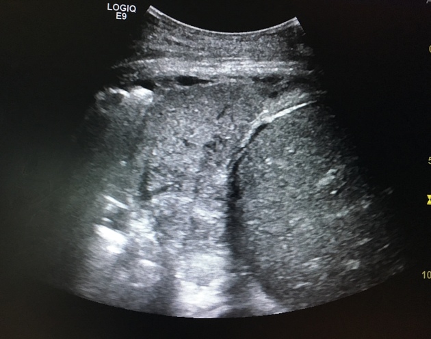
The diapgragmatic slip delineates the liver from the dense lower lobe consolidation. The lung appears nearly identical to the liver in terms of density.
One can also well illustrate that the appearances on the chest radiograph do not relate to fluid.
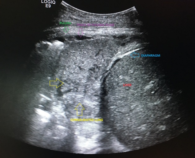
Annotations indicate the key features from the focused ultrasond of the chest.
Case Discussion
Ultrasound is very helpful, infact the gold standard imaging investigation, for assessing the content of the pleural space and the assessment of pleural fluid versus consolidated lung.
This case illustrates how effective ultrasound of the chest is for assessment of pleural fluid versus other causes for radiographic appearances.
Furthermore CT may depict the thoracic cavity including pleura, but it is less sensitive for the assessment of the internal content of pleural fluid, such as viscosity, loculations and septations. Both are important in management decisions with complex pleural collections requiring VATS, as opposed to radiological percutaneous drain insertion for simple collections.


 Unable to process the form. Check for errors and try again.