
Presentation
Two year history of swelling on the volar aspect of the index finger. Giant cell tumor of tendon sheath?
Patient Data
Age: 30 years
Gender: Female
From the case:
Giant cell tumor of tendon sheath


Download
Info

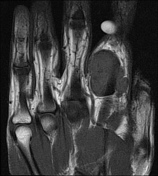
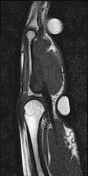
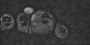
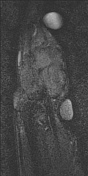
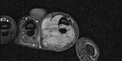
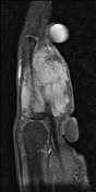
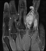
Skin surface markers in situ.
Well defined, lobulated 3.5x2.2x2.1cm T1 isointense, heterogeneously enhancing mass seen around the index finger metacarpal.
The lesion encases the flexor tendon with scalloping of the metacarpal base. It's predominantly on the palmar surface and extending dorsally with the main bulk on medial aspect.
Case Discussion
The appearances of this peritendinous mass on the index finger are most consistent with a giant cell tumor of the tendon sheath.
To date, surgery has not been performed for a confirmed histological diagnosis.


 Unable to process the form. Check for errors and try again.